Call us now
07971459752
Human Ear Model

Human Ear Model Specification
- Features
- Detachable parts, Enlarged for clarity, Color-coded
- Temperature Resistance
- Standard classroom conditions
- Control Type
- Manual
- Display Type
- Three-dimensional physical model
- Accuracy
- Realistic anatomical representation
- Shape
- Three-Dimensional Model of Human Ear
- Type
- Anatomical Model
- Dimension (L*W*H)
- Approx. 12 cm x 7 cm x 4 cm
- Equipment Type
- Human Ear Model
- Equipment Materials
- Plastic
- Material
- PVC
- Application
- Medical Education, Demonstration, Anatomy Study
- Finish
- Smooth, Painted Finish
- Usage
- Educational Demonstration, Biology Labs, Anatomy Teaching
- Weight
- Approx. 300 g
- Detachable Parts
- Yes
- Recommended Users
- Medical Students, Teachers, Educational Institutes
- Parts Included
- Outer, Middle, and Inner Ear Sections
- Mounting
- Mounted on a Base Stand
- Color
- Multi-color for part differentiation
- Cleaning Instructions
- Wipe with Damp Cloth
About Human Ear Model
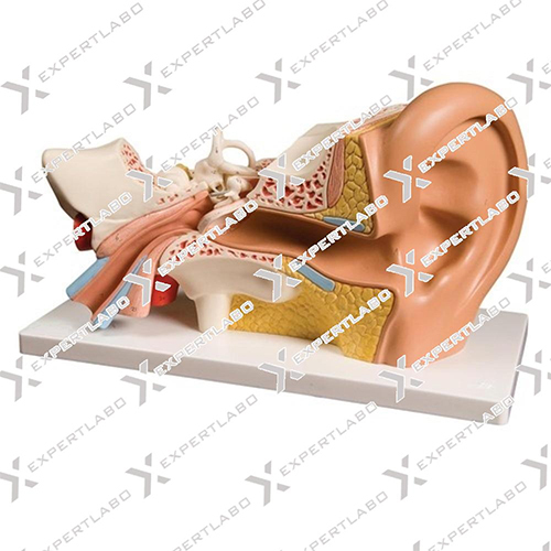
3X life size. The model shows details of the external, middle and inner ear. The eardrum with malleus, incus and stapes are removable. The other removable part is composed of the cochlea and labyrinth with vestibular and cochlear nerves.Detailed, Color-Coded Anatomy
This anatomical model carefully presents the main components of the human ear-outer, middle, and inner sections-with each part color-coded for easy identification. The smooth, painted finish enables students and teachers to visually distinguish key structures, greatly aiding the understanding of complex ear anatomy.
Ideal for Teaching and Demonstrations
With detachable sections mounted on a sturdy base, this model is perfect for hands-on educational use. Designed for medical students, teachers, and academic institutions, it supports interactive teaching and demonstration in biology labs, classrooms, and anatomy studies, making learning more engaging and effective.
Durable and Easy to Maintain
Manufactured from high-quality PVC and plastic, this model withstands regular classroom use under standard conditions. It is lightweight, portable, and simple to maintain-requiring only a damp cloth for cleaning. The design ensures longevity and safe handling during repeated demonstrations.
FAQ's of Human Ear Model:
Q: How are the different parts of the ear model distinguished for study?
A: Each section of the model-outer, middle, and inner ear-is color-coded and labeled, allowing users to easily identify and differentiate anatomical parts during demonstrations.Q: What is the intended usage of the human ear model?
A: This model is designed for educational demonstrations, biology labs, anatomy teaching, and medical training, providing interactive and visual support for understanding ear structures.Q: When should the detachable parts be used during instruction?
A: Detachable parts are best utilized when illustrating specific structures or processes within the ear. Students and instructors can disassemble the model to examine internal components closely during detailed lessons.Q: Where is the model best utilized for demonstrations and teaching?
A: The model is ideal for use in classrooms, biology laboratories, anatomy courses, and other educational settings in medical schools and institutes.Q: What is the cleaning and maintenance process for the ear model?
A: To clean the model, simply wipe it gently with a damp cloth. Avoid using harsh chemicals to preserve the painted, smooth finish and the clarity of the color-coded sections.Q: What are the primary benefits of using this anatomical ear model in an educational setting?
A: The model enhances comprehension through three-dimensional visualization, color-coding, and interactive components. It supports experiential learning for students studying human anatomy, especially the auditory system.Tell us about your requirement

Price:
Quantity
Select Unit
- 50
- 100
- 200
- 250
- 500
- 1000+
Additional detail
Mobile number
Email

More Products in Biology Models Category
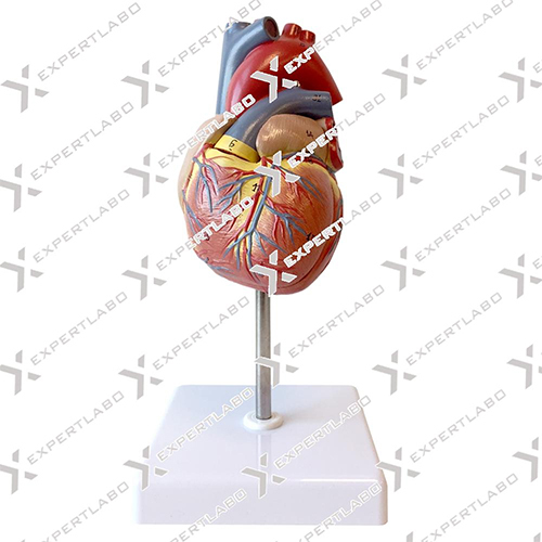
Human Heart
Price 1000 INR / Unit
Minimum Order Quantity : 1 Unit
Equipment Materials : PVC Plastic
Display Type : Physical 3D Model
Material : Polyvinyl Chloride (PVC)
Features : Dissectible, Colored Structures, ReAssemblable
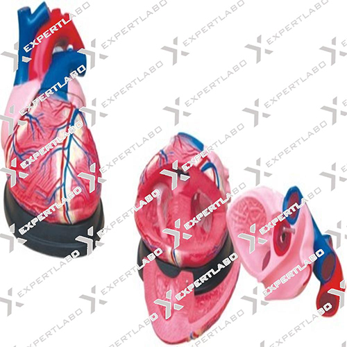
Jumbo Heart Model
Price 1800 INR
Minimum Order Quantity : 1 Unit
Equipment Materials : PVC Plastic
Display Type : 3D Physical Model
Material : Nontoxic, durable PVC
Features : Detachable parts, Handpainted Details, Enlarged for Ease of Study
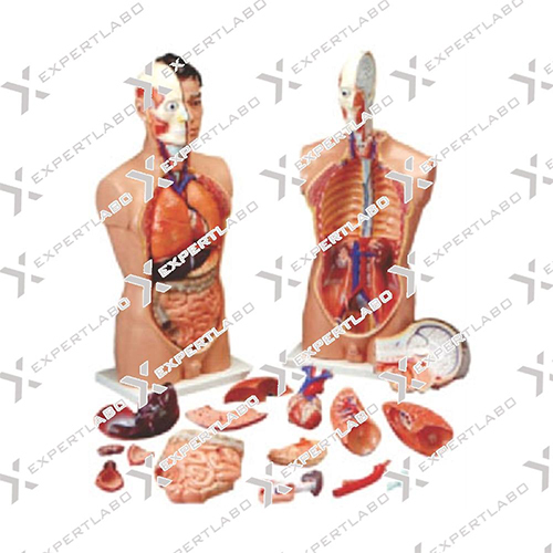
Human Anatomy Torso 18 Parts
Price 5000 INR / Unit
Minimum Order Quantity : 1 Unit
Equipment Materials : PVC Plastic
Display Type : Physical 3D Model
Material : HighQuality Durable PVC
Features : 18 Parts, Disassemblable, Colored for Clarity
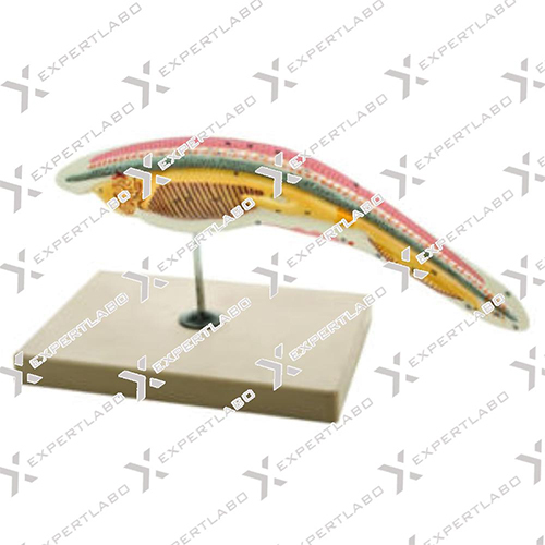
Amphioxus Models
Price 8000 INR
Minimum Order Quantity : 1 Unit
Equipment Materials : Plastic, PVC
Display Type : 3D Physical Model
Material : HighQuality Durable PVC Plastic
Features : Hand Painted, Enlarged Detailing, Mounted on Base, NonToxic
Expert Labo
GST : 06BELPA5037D2ZX
GST : 06BELPA5037D2ZX
Office 1001, Housing Board Colony, Ambala Cantt, Ambala Cantt - 133001, Haryana, India
Phone :07971459752
Mr Ajay Manocha
(Proprietor)
Mobile :07971459752
Mr- Ajay Manocha
(Proprietor)
Mobile :07971459752
- Biology Models
- Laboratory Equipment
- Pharmaceutical Lab Equipment
- Laboratory Instruments
- Physics Equipments
- Laboratory Glassware
- Laboratory Apparatus
- Hospital Equipment
- Physics Instruments
- Testing Machine
- Hospital Furniture
- Oxygen Flow Meter
- Educational Aids
- Biological Models
- Hospital Bed
- Cement Testing Equipment
- Soil Testing Equipment
- Anatomical Models
- Suction Machine
- Hospital Uniform
- Autoclave And Sterilizer
- Mortuary Cabinet
- Laboratory Microscope
- Medicine Cabinet
- Crash Cart Trolley
- Surgical Tray
- Compression Testing Machine
- Hot Air Oven
- Lab Safety Products
- Laboratory Testing And Measuring Instrument
- Survey Total Station
- Blood Pressure Monitor
- Dialysis Chair
- Examination Couches
- OT Lights
- OT Tables
- Patient Transfer and Handling System
- Ward -OT Equipments
- Examination Lights
 Send Inquiry
 Send SMS
Expert Labo
All Rights Reserved.(Terms of Use)
Developed and Managed by Infocom Network Private Limited.
Developed and Managed by Infocom Network Private Limited.

