Call us now
07971459752
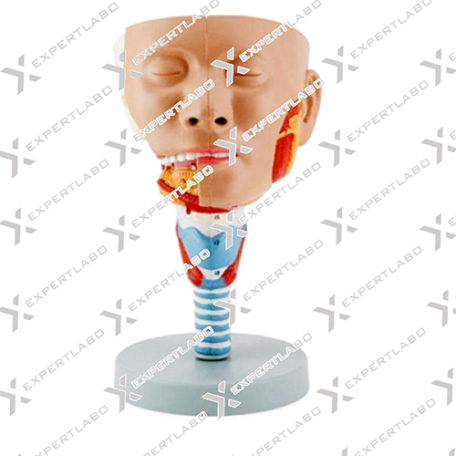
Head With Pharynx Muscles

Head With Pharynx Muscles Specification
- Features
- Detachable Parts, Colored Details, Numbered Structures
- Control Type
- Manual Display
- Temperature Resistance
- Up to 50C
- Shape
- Head with Neck and Pharynx
- Accuracy
- High Anatomical Accuracy
- Display Type
- 3D Physical Model
- Type
- Anatomical Model
- Dimension (L*W*H)
- Life-size (Approx. 20 x 15 x 28 cm)
- Equipment Type
- Demonstration Model
- Equipment Materials
- PVC Plastic
- Material
- Premium Quality PVC
- Application
- Medical Education, Anatomy Study
- Number of Parts
- 3 Parts, Separately Removable
- Structure Details
- Depicts Deep Muscular, Nervous, and Vascular Structures of the Pharynx and Associated Regions
- Intended Users
- Medical Students, Instructors, Professionals
- 3D Visualization
- Realistic Representation
- Mobility
- Portable
- Compatibility
- Suitable for Classroom and Laboratory Use
- Color Coding
- Hand-painted for Easy Identification
- Mounting
- Mounted on Base Stand
- Cleaning Instructions
- Wipe Clean with Damp Cloth
About Head With Pharynx Muscles
This life size head model is dissected along the sagittal plane into 2 halves. Details of the oronasal cavity and larynx as well as musculature of the pharynx are exceptionally well represented. Mounted on base with stand.
Highly Detailed Educational Model
The Head With Pharynx Muscles model provides an advanced depiction of deep muscular, nervous, and vascular structures, enhancing the learning experience for anatomy students and educators. Its life-size dimensions and accurate structure allow for in-depth study in a variety of educational environments.
Interactive and Durable Design
Composed of premium quality PVC, this anatomical model offers durability while being lightweight and portable. The three detachable parts allow users to explore complex anatomical features easily, making it an ideal demonstration tool for practical teaching sessions.
Color-Coded Clarity and Easy Maintenance
Hand-painted with vibrant color coding, each structure on the model is easy to identify and numbered for reference. Maintenance is simple-just wipe it clean with a damp cloth. The model withstands temperatures up to 50C, ensuring longevity in busy classroom or laboratory settings.
FAQ's of Head With Pharynx Muscles:
Q: How can the Head With Pharynx Muscles model enhance anatomy education?
A: The model offers a realistic, life-size representation of the pharynx and related structures, with color-coded, hand-painted details, making it easier for students and educators to identify, study, and understand deep anatomical relationships.Q: What are the different parts included in this anatomical model?
A: The model consists of three separately removable sections that describe deep muscular, nervous, and vascular structures, enabling detailed analysis from different perspectives.Q: When is it most beneficial to use this model?
A: This model proves most useful during classroom lessons, laboratory sessions, practical demonstrations, and during preparation for exams or clinical practice, as it helps reinforce theoretical concepts with a physical 3D reference.Q: Where can this anatomical model be effectively utilized?
A: It is suitable for use in medical schools, teaching hospitals, university laboratories, and anatomy classrooms, offering educational value wherever hands-on anatomical study is important.Q: What process is followed for cleaning and maintaining the model?
A: Cleaning the model is simple-just use a damp cloth to wipe the surfaces. The premium PVC material ensures resilience and resistance to classroom temperature variations up to 50C.Q: Who are the intended users of this demonstration model?
A: This model is designed for medical students, anatomy instructors, and healthcare professionals involved in medical education and anatomy studies.Q: Why is the hand-painted, color-coded feature beneficial?
A: Color-coding and hand-painting facilitate quick identification of anatomical features, aiding visual memory and improving comprehension during both self-study and group teaching sessions.Tell us about your requirement

Price:
Quantity
Select Unit
- 50
- 100
- 200
- 250
- 500
- 1000+
Additional detail
Mobile number
Email

More Products in Biology Models Category
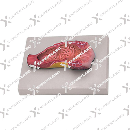
Human Anus Model
Price 5000 INR / Unit
Minimum Order Quantity : 1 Unit
Dimension (L*W*H) : Approx. 20 cm x 20 cm x 15 cm
Shape : Anus and Surrounding Tissue
Application : Medical Training, Patient Education, Classroom Demonstration
Equipment Type : Anatomical Model
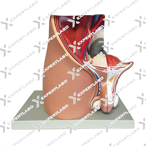
Male Pelvis 4 Parts
Price 2500 INR
Minimum Order Quantity : 1 Unit
Dimension (L*W*H) : Approx. 30 x 18 x 20 cm
Shape : Threedimensional cross sectional model
Application : Medical Education, Demonstration, Anatomy Study
Equipment Type : Human Anatomical Model
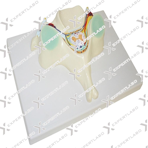
5TH Cervical Vertebra
Price 5200 INR
Minimum Order Quantity : 1 Unit
Dimension (L*W*H) : Approx. 8 cm x 7 cm x 4 cm
Shape : Fifth Cervical Vertebra (C5)
Application : Medical Education, Anatomy Study
Equipment Type : Human Vertebrae Model
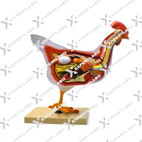
Hen Dissection Model
Price 2000 INR / Unit
Minimum Order Quantity : 1 Unit
Dimension (L*W*H) : Approx. 38 cm x 25 cm x 6 cm
Shape : Rectangular
Application : Biology, Zoology, Educational Purposes, Dissection Study
Equipment Type : Anatomical Model
Expert Labo
GST : 06BELPA5037D2ZX
GST : 06BELPA5037D2ZX
Office 1001, Housing Board Colony, Ambala Cantt, Ambala Cantt - 133001, Haryana, India
Phone :07971459752
Mr Ajay Manocha
(Proprietor)
Mobile :07971459752
Mr- Ajay Manocha
(Proprietor)
Mobile :07971459752
- Biology Models
- Laboratory Equipment
- Pharmaceutical Lab Equipment
- Laboratory Instruments
- Physics Equipments
- Laboratory Glassware
- Laboratory Apparatus
- Hospital Equipment
- Physics Instruments
- Testing Machine
- Hospital Furniture
- Oxygen Flow Meter
- Educational Aids
- Biological Models
- Hospital Bed
- Cement Testing Equipment
- Soil Testing Equipment
- Anatomical Models
- Suction Machine
- Hospital Uniform
- Autoclave And Sterilizer
- Mortuary Cabinet
- Laboratory Microscope
- Medicine Cabinet
- Crash Cart Trolley
- Surgical Tray
- Compression Testing Machine
- Hot Air Oven
- Lab Safety Products
- Laboratory Testing And Measuring Instrument
- Survey Total Station
- Blood Pressure Monitor
- Dialysis Chair
- Examination Couches
- OT Lights
- OT Tables
- Patient Transfer and Handling System
- Ward -OT Equipments
- Examination Lights
 Send Inquiry
 Send SMS
Expert Labo
All Rights Reserved.(Terms of Use)
Developed and Managed by Infocom Network Private Limited.
Developed and Managed by Infocom Network Private Limited.

